Non-Invasive Scanning and Subtle Energy Testing Lab
MEMO OF UNDERSTANDING
Effects of Biophotonic Light on Acute Inflammation as seen with Thermal Imaging: Preliminary study
Principal Investigator: Gaétan Chevalier, Ph.D., Research Director (Bio in Appendix A)
Co-Investigator: Mary D. Clark, Ph.D., Managing Director (Bio in Appendix B)
Goal: This is a preliminary project using thermal imaging (TI; see appendix C) to assess the effect of Biophotonic Light on acute inflammation.
Statement of Work:
One (1) subject with acute inflammation and pain of the left knee was recruited to participate in this project. Two thermal images were taken. A thermal image was taken before application of the Biophotonic Light and another one 10 minutes after. The VAS pain scale was used to determine her level of paint.
Results:
Thermal images:

VAS Pain Scale:
In this scale 0 means no pain and 10 is the most imaginable pain possible. This is for her left knee, the knee with most pain.
The subject reported feeling slightly better after having 10 minutes of Biophotonic Light therapy compared to when she arrived in the lab. Her comment was that she felt warmer from the light and that was comforting and soothing and that her left knee felt better than the right knee which was the opposite of the situation she was in when she arrived..
Conclusion:
Ten (10) minutes application of Biophotonic Light application resulted in a significant difference in thermal images before and after Biophotonic Light application. Also the subject reported a significant decrease in pain. This result warrants a larger study to confirm these preliminary results.
APPENDIX A
Gaétan Chevalier, Ph.D.,
Biographical Sketch
Dr. Gaétan Chevalier received his Ph.D. from the University of Montréal in Atomic Physics and Laser Spectroscopy in 1988. After 4 years of research at UCLA in the field of nuclear fusion, he became professor and Director of Research at the California Institute for Human Science (CIHS) in 1993 where, for 10 years, he conducted research projects on human physiology and electrophysiology as well as being Director of the Life Physics Department and Research Director. Dr. Chevalier is currently Research Faculty at CIHS, and he has been Director of Research at Psy-Tek Labs since June 2010.
APPENDIX B
Mary D. Clark, Ph.D.,
Biographical Sketch
Mary D. Clark, Ph.D. is a licensed psychologist in Arizona, and is a licensed marriage family therapist and licensed educational psychologist in California. She maintains both a private practice and a healing practice in Encinitas, California. Mary is a Certified Energy Healing Instructor, a Senior Certified Energy Healer, and past coordinator of the Energy Healing Certification Program for the central and western states. She has practiced and taught Energy Healing for over 20 years.
APPENDIX C
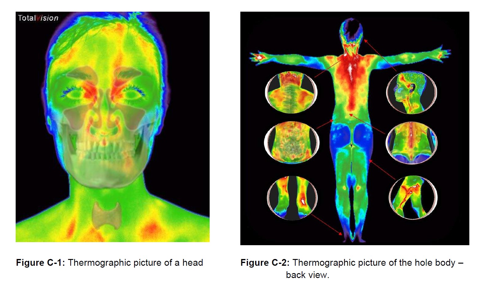
Medical Thermal Imaging
Thermography is an American invention initially used in World War II as a method of tracking troops and movement in the dark and at great distances. As a military device, it was highly classified until the 1960’s, when it was officially declassified and became available for other uses, although it is still used extensively in military and other government projects.
Today, thermography is used extensively industrially and in medicine. After declassification, the National Cancer Institute accepted the use of thermography for breast imaging until mammography became the focus of breast imaging. Consequently, radiologists embraced mammography as a radiological device since it fell within their specialty and was responsible for advancements for doctors within that specialty. Together with the efforts of the radiologists to eliminate the competition and the lack of understanding of thermography combined with the lack of knowledge of the physiological sympathetic and autonomic responses of the body, thermography was effectively pushed aside. Coupled with the public’s demand for safe imaging and some pioneers in the fields of medicine and industrial business thermography has been brought back to the mainstream.
Medical Thermal Imaging is the use of an infrared camera to “see” and “measure” what we call metabolism, thermoregulation or thermal energy emitted from the body. Thermography reveals a fascinating and reliable pattern of thermal activity that discloses a silent warning. A sensitive infrared video camera can even detect a gentle but visible pulsation created by blood pumping through blood vessels.
Infrared lets us see what our human eyes cannot; because infrared is part of an invisible electromagnetic wavelength that is perceived as heat. Everything with a temperature above absolute zero emits heat. Infrared thermography cameras provide the ability to record precise non-contact body temperature measurement. Abnormal physiological activities in the body are easily observed with thermography.
Thermography is not a stand-alone diagnostic device and does not replace any other diagnostic device or examination and is therefore used as an adjunctive screening tool with other such devices and examinations.

